
Light microscopy is often the first step in the analysis of solid samples. A modern light microscope is not just a very good “magnifying glass” but provides insight into the structural and chemical heterogeneity of materials based on different image contrasts.
Bright field and dark field
In normal bright-field microscopy, light is focused by an objective lens (characterized by its magnification and numerical aperture (NA), e.g. 50x/NA = 0.75) onto the sample and the light either reflected (reflected light microscopy) or transmitted (transmitted light microscopy) by the sample is collected. The image contrast is primarily based on light absorption.
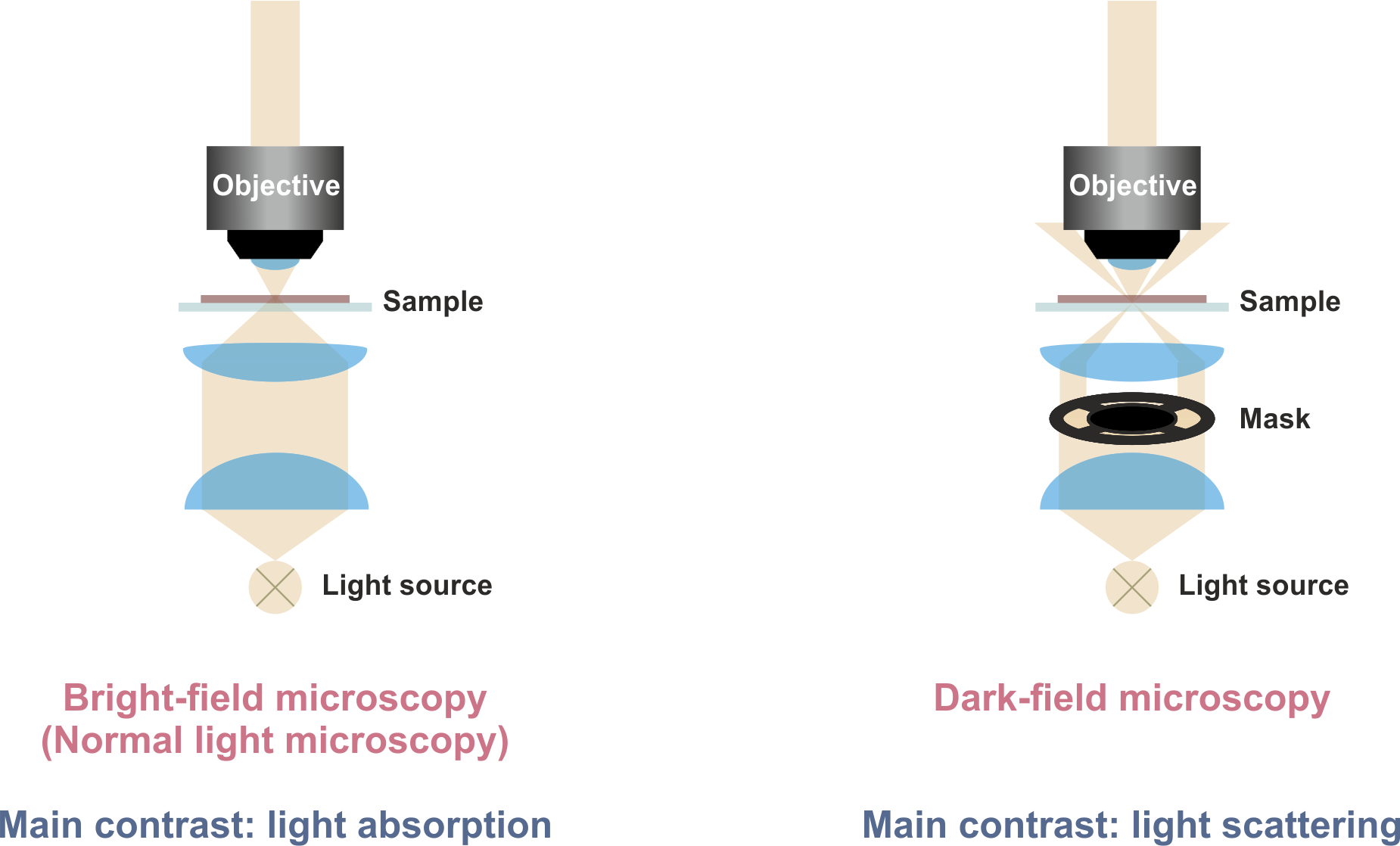
In dark-field microscopy, the inner part of the illuminating light beam is blocked by a mask. Thus, only a sheet of light is focused onto the sample. In other words, the sample is illuminated under an angle that is defined by the NA of the objective lens. Only the fraction of light is detected, which has changed its angle upon interaction with the sample. The image contrast is primarily based on light scattering, enhancing the contrast of, e.g., small particles and sharp edges.
Polarized light microscopy
In polarized light microscopy, two polarization filters are introduced into the beam path. The polarizer converts white light (as a mixture of all polarization directions) into linearly polarized light.
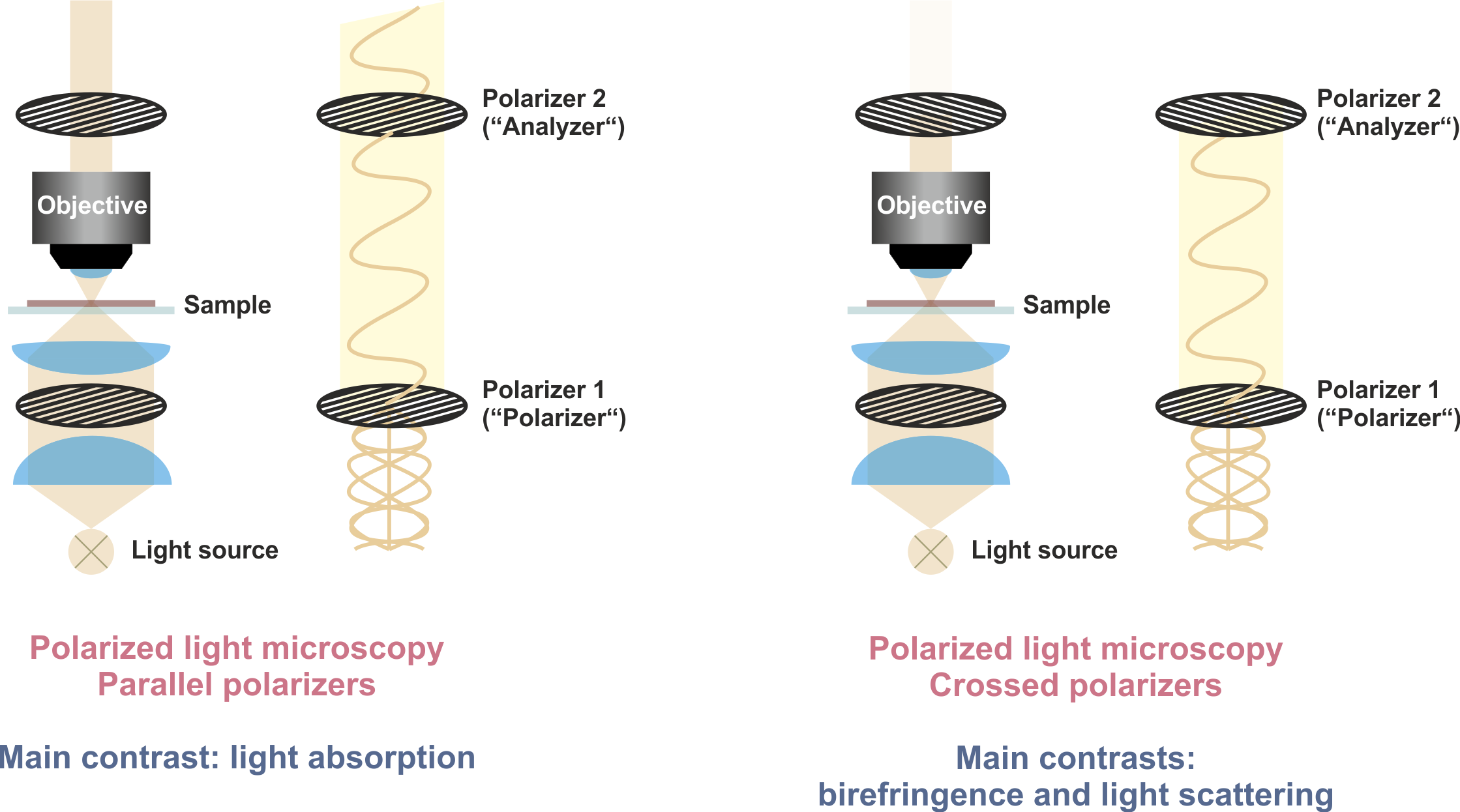
If an analyzer with 90° orientation with respect to the polarizer is used (crossed polarizers), only light can reach the camera, which has changed its polarization direction upon interaction with the sample. In this image contrast, amorphous parts of the sample appear dark, whereas the contrast of, e.g., certain crystals and small particles is enhanced, which can change the light polarization due to birefringence or light scattering, respectively.
 ORCID 0000-0001-9708-931X