
Spectroscopic imaging
An image is worth a thousand spectra
![]()
Microscopes enable the look into the fascinating world of the micrometer and nanometer scales, which is hidden to the naked eye. When looking at complex samples, such as biological cells or micro and nano-structured materials, questions about what exactly is seen on an image arise not only to the layman, because information on chemical compositions and physical properties is missing. Spectroscopic imaging methods deal with this issue by allowing to add “chemical sense” to microscope images.
Such methods, also termed microspectroscopy, are based on spectroscopic techniques, which analyze matter based on its interaction with electromagnetic radiation (e.g. visible light, X-rays etc.). Intensity distributions of the various wavelengths (simply called ‘spectra’) that are characteristic for individual sample constituents enable the identification of atoms, molecules or crystal structures (qualitative analysis) and in many cases also the measurement of their concentration or amount (quantitative analysis).
Microspectroscopy combines this analytical approach with microscopy. Sample surfaces are raster-scanned by a microscopic measurement volume, and in each measurement spot (that is, each pixel of an image) a full spectroscopic measurement is performed. From this data, distributions of, e.g., chemical structures can be calculated.
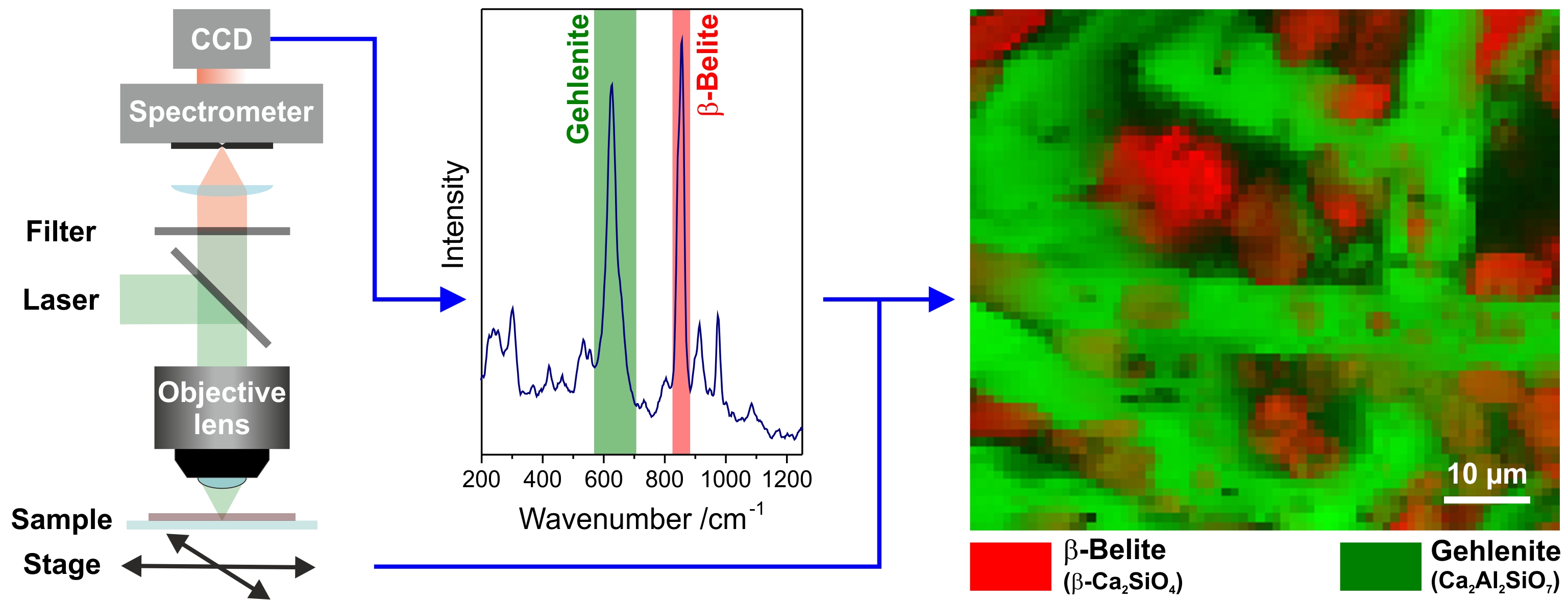
Principle of Raman microscopy: A sample is raster-scanned with a laser beam, and in each pixel a spectrum (here: Intensity vs. Wavenumber) is acquired, which contains signal patterns that are characteristic for the various sample constituents. Analysis of selected signal intensities yields distribution maps of chemical compounds (here: inorganic phases in a 19th-century cement stone sample). (see: Petra Dariz, Thomas Schmid, in: Mater. Charact. 129 (2017) 2.*)
Each microspectroscopic technique is basically a “chemical eye” enabling the look at a certain part of the sample properties. For example, elemental analysis techniques look at atoms and provide distributions of the elements of the periodic table (e.g. scanning electron microscopy with energy dispersive X-ray spectroscopy, SEM-EDX), whereas molecular spectroscopy techniques analyze chemical bonds and thus, provide distributions of molecules and crystals (e.g. Raman microscopy). Complex analytical questions often can only be solved by combining such complementary information.
The look at the microstructure and chemical composition of materials provided by spectroscopic or chemical images, respectively, is straightforward and contributes to a deeper understanding of their properties and function. Therefore, a 100 x 100-pixel image is really worth the acquisition and analysis of 100 x 100 = 10’000 individual spectra.
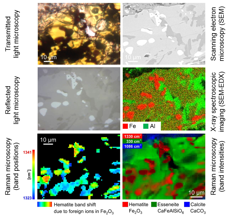
Different „chemical eyes“ reveal different properties of a material sample: Elemental analysis via SEM-EDX looks at the different chemical elements (here iron and aluminium), whereas molecular spectroscopy in the form of Raman microscopy visualizes some of the chemical compounds and even provides insight into small variations within crystal lattices (here: the local implementation of foreign ions into hematite α-Fe2O3). (see: Petra Dariz, Thomas Schmid, in: Mater. Charact. 129 (2017) 2.*)
* The figures in this section are adapted and extended from:
Materials Characterization
 |
P. Dariz, T. Schmid Ferruginous phases in 19th century lime and cement mortars: A Raman microspectroscopic study Mater. Charact. 129 (2017) 2–17 |
| PDF © | Abstract (free) Supplementary material (free) |
Research
My current research projects are best summarized by the following keywords:
- Analytical Chemistry/Analytical Sciences
- application-related
- interdisciplinary
- Chemical/spectroscopic imaging
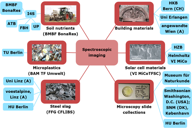
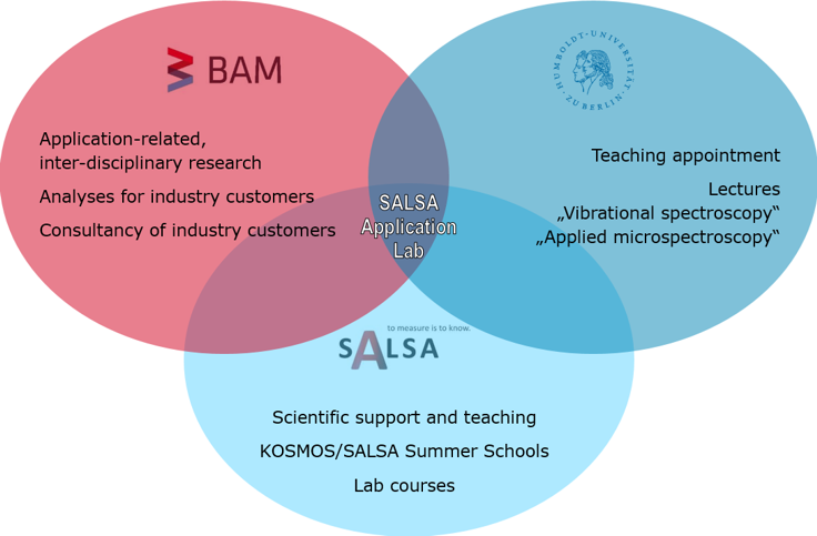
Most often, I work with collaboration partners from various disciplines (with Analytical Sciences connecting them) in application-related research projects. By using chemical/spectroscopic imaging (with a focus on Raman microscopy) we aim to solve questions, which can only be solved in a real interdisciplinary collaboration, in which the sum is greater than the parts and the parts are open-minded to learn from each other (external collaboration partners: see blue rectangles in the figure).
Current research questions:
- Phase analysis and determination of historical mortars, including 19th-century cements, high-fired medieval gypsum
- Analysis of thin-film solar cell materials such as Cu(In,Ga)Se2 (phase analysis, crystal orientations, stress, strain)
- Determination of embedding media of microscope slides from natural history collections and investigation of their aging processes
- Element and phase analyses of steel slags
- Detection of microplastics in environmental matrices
- Spatially resolved determination of soil nutrients (precision farming)
(Research: see also Publications)
Analyses for industry customers
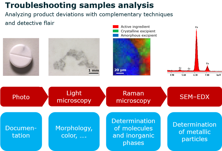
Knowledge and experiences gained in research projects often find their way to applications in the „real world“. We provide in-depth analyses of product deviations to industry customers of the Federal Institute for Materials Research and Testing (BAM). This ‚troubleshooting samples analysis‘ focuses on (without being limited to) particular contaminants in solid and liquid samples at the micrometer to millimiter size scales. We aim to gather fast and reliable answers about their possible sources and reasons.
A typical example are local color changes in tabletted products. As most often it is unknown a priori, whether the contaminants are metallic, inorganic, or organic, analytical techniques complementing each other need to be employed (see figure below).
Futhermore, in the frame of the consortial study ‚InLight‘ we informed industry customers about new trends in process analysis and discussed with them further possibilities and needs in process analytical technology.
HU Berlin and SALSA
The School of Analytical Sciences Adlershof (SALSA) is a graduate school at Humboldt-Universität zu Berlin (HU) enabling PhD students from all over the world to perform their PhD projects in an interdisciplinary environment and to widen their knowledge of Analytical Sciences in seminars, lectures, summer schools and lab courses. Further to teaching appointments at HU regarding undergraduate classes in Chemistry, my work for HU includes the setup and administration of the SALSA Application Lab (see below) as well as scientific support, teaching and co-organization of summer schools at SALSA.
(teaching: see also Vorlesungen/Lectures)
- Lab course SALSA: „Applied Microspectroscopy“
- Teaching appointment HU: Lecture „Schwingungsspektroskopie (IR/Raman)“/“Vibrational spectroscopy (IR/Raman)“, BSc class in Chemistry
- Lecture HU: „Angewandte Mikrospektroskopie“/“Applied Microspectroscopy“ in the advanced lecture series „Analytische Chemie“/“Analytical Chemistry“, MSc class in Chemistry
- KOSMOS Summer University 2014 „Limits and Scales in Analytical Sciences“
- SALSA Summer University 2015 „Sensitivity and Selectivity in Analytical Sciences“
- SALSA Summer University 2016 „Make and Measure in Analytical Sciences“
SALSA Application Lab
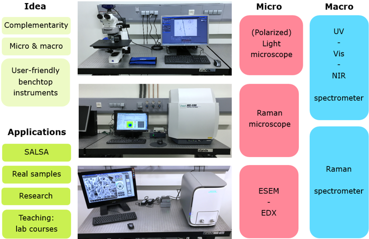
Last but not least, in 2015/2016 I was enabled to equip the SALSA Application Lab based on my experiences from application (analyses for industry) and application-related research. As lab supervisor, I use the application lab for
- SALSA teaching (e.g. lab courses)
- Research projects of SALSA fellows
- application-related research
- real samples
- research collaborations with colleagues from SALSA, BAM, companies, ….
The SALSA Application Lab offers the following analytical techniques (with a focus on spectroscopy and imaging):
 ORCID 0000-0001-9708-931X